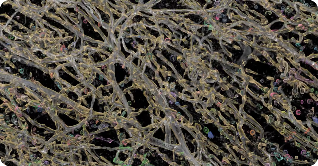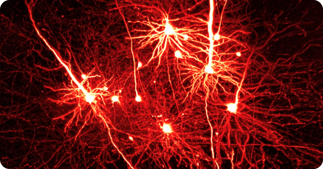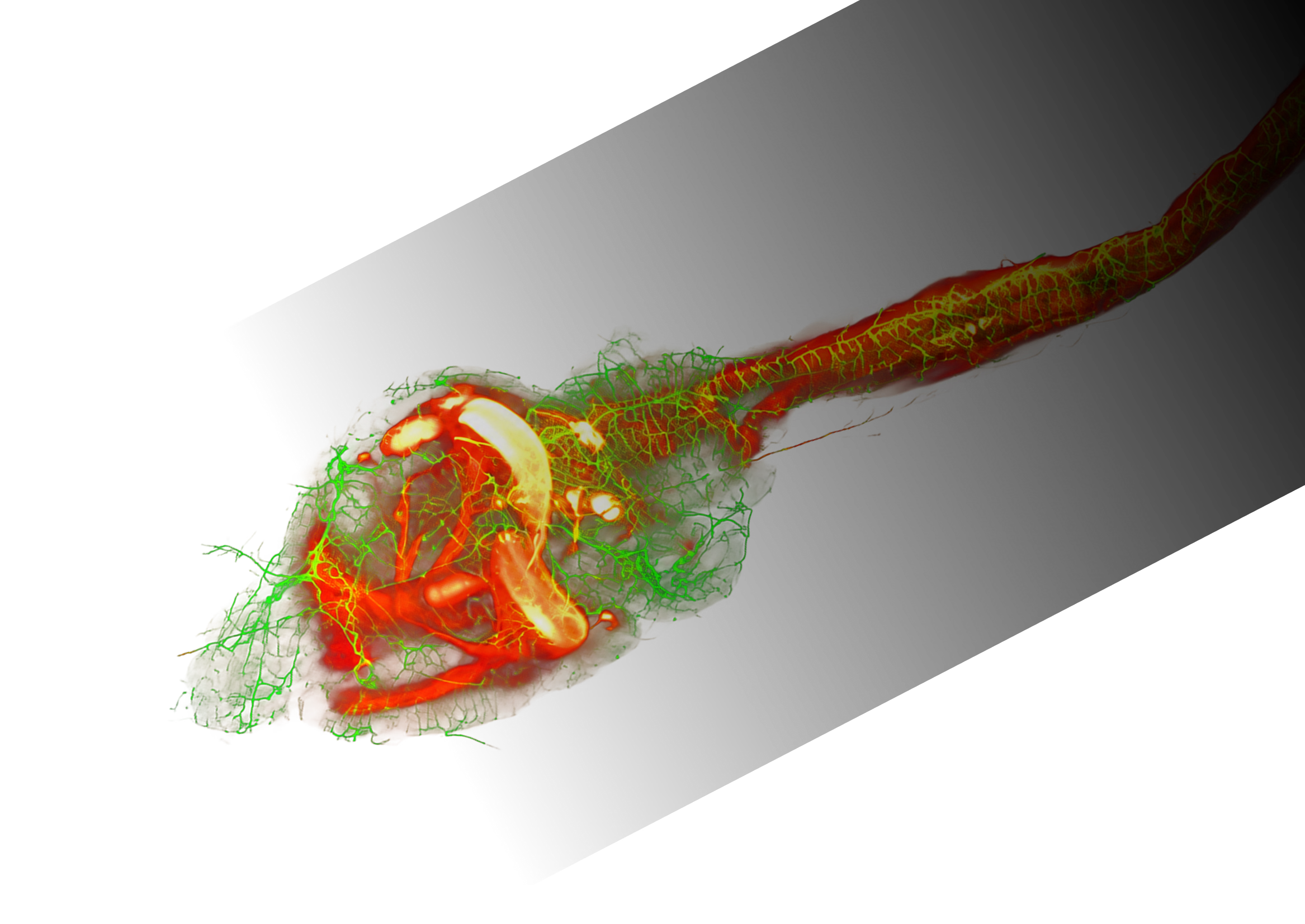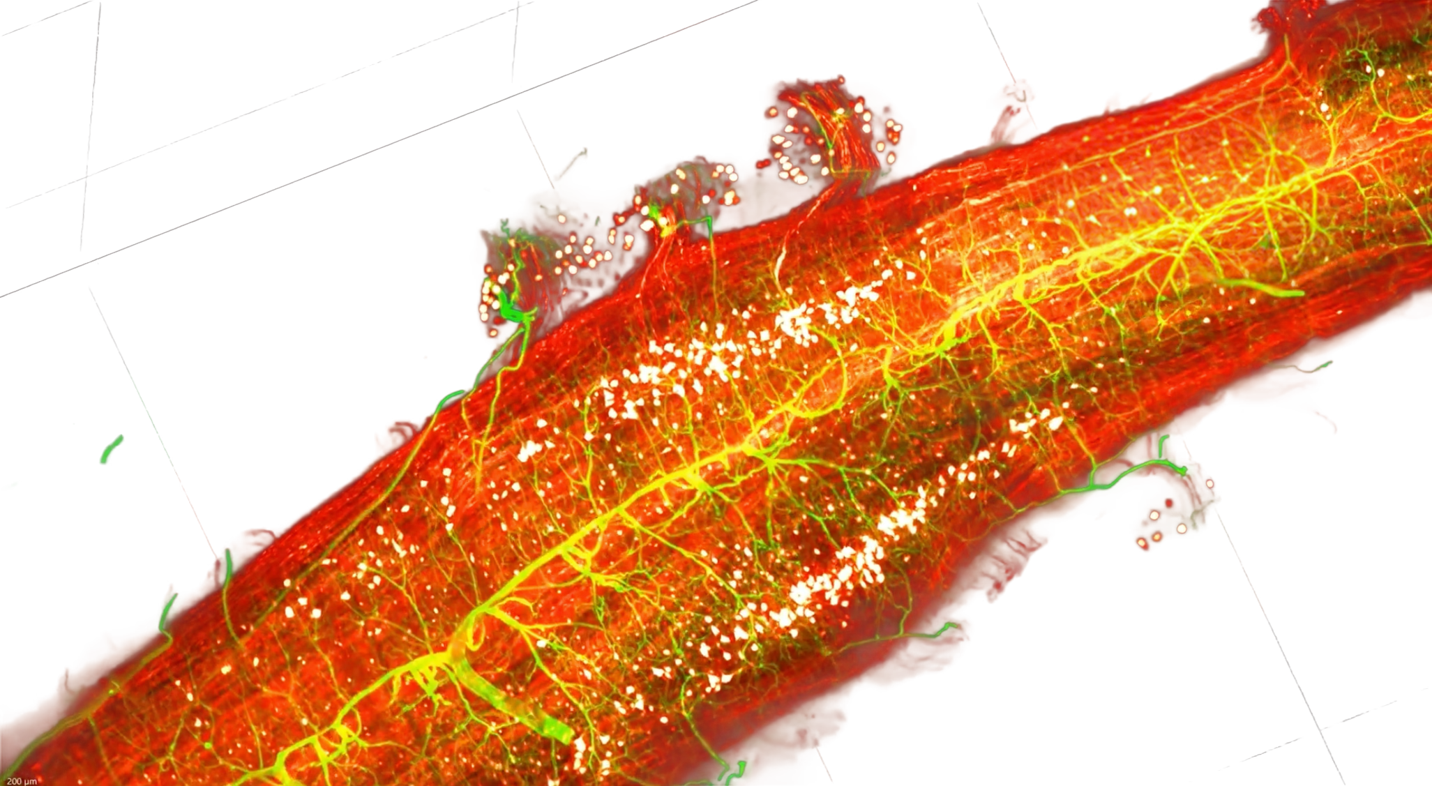
High-res imaging of intact cleared tissue

XL2 combines high-resolution volumetric imaging with flexible sample handling and an integrated acquisition-to-analysis workflow.

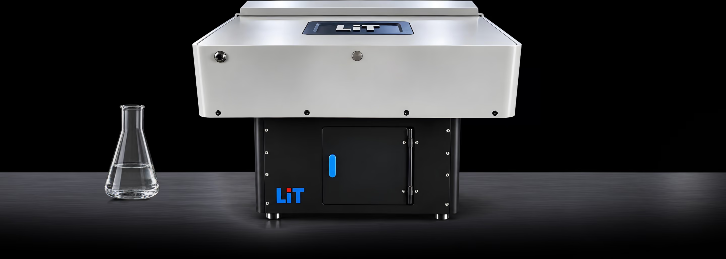
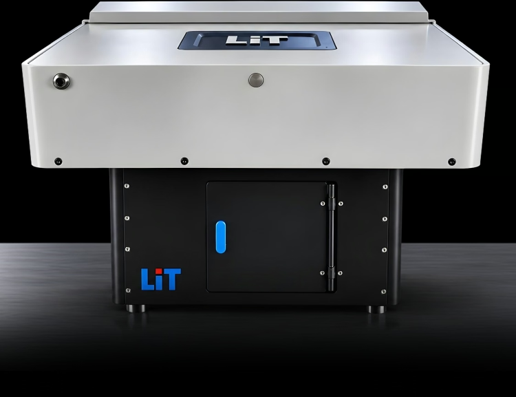
LiT XL2 microscope on a laboratory benchLiT XL2 microscope on a laboratory bench

Detail, consistency and flexibility in one workflow

High-resolution volumetric imaging

Resolve fine structures across intact cleared tissues without physical sectioning.

Uniform imaging across dense samples

More consistent illumination with fewer dark regions and less striping.

Flexible sample handling

Adapt to different specimens, clearing methods, immersion media and imaging scales.

Integrated workflow

Move from acquisition and processing to 3D rendering and downstream analysis.

The optics behind the image

LiT was founded around patented Line Bessel Sheet technology. XL2 combines it with four-side illumination, refractive-index correction and a range of objectives.

Line Bessel Sheet
Long, thin light sheets for high-resolution imaging with reduced side-lobe background
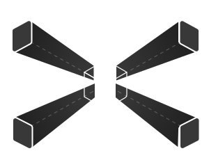
Four-side illumination
More uniform illumination across dense cleared samples with less striping and shadowing
RI correction optics
RI 1.33–1.58
Adjustable refractive-index correction from 1.33 to 1.58 for different clearing media
Multiple objectives
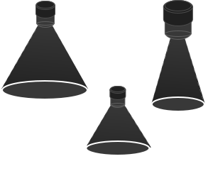
4×5×10×16×25×
Flexible magnification from whole-sample overview to fine structural detail
Proven in practice

Demonstrated on intact tissue, organs and whole organisms

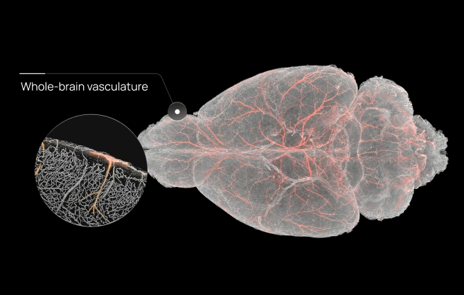
Whole brain macro- and micro-vasculature at sub-micron resolution

Vasculature
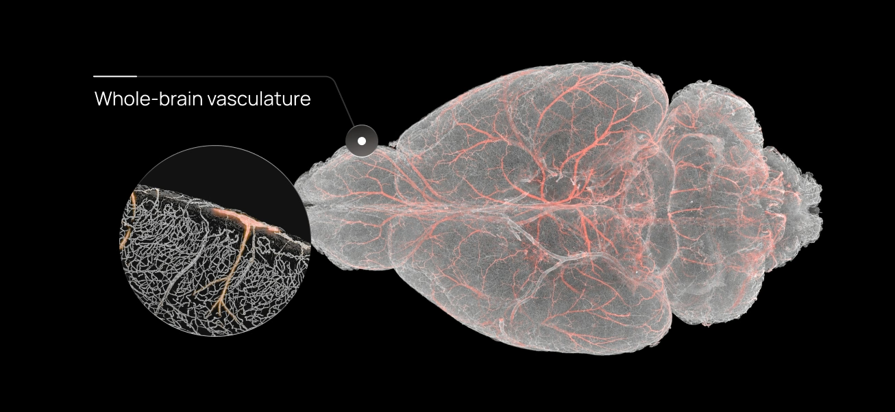
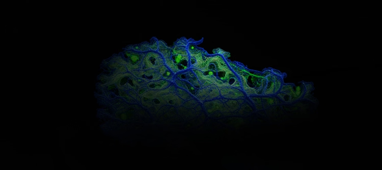
Whole-brain vasculature with magnified detailWhole-brain vasculature with magnified detail

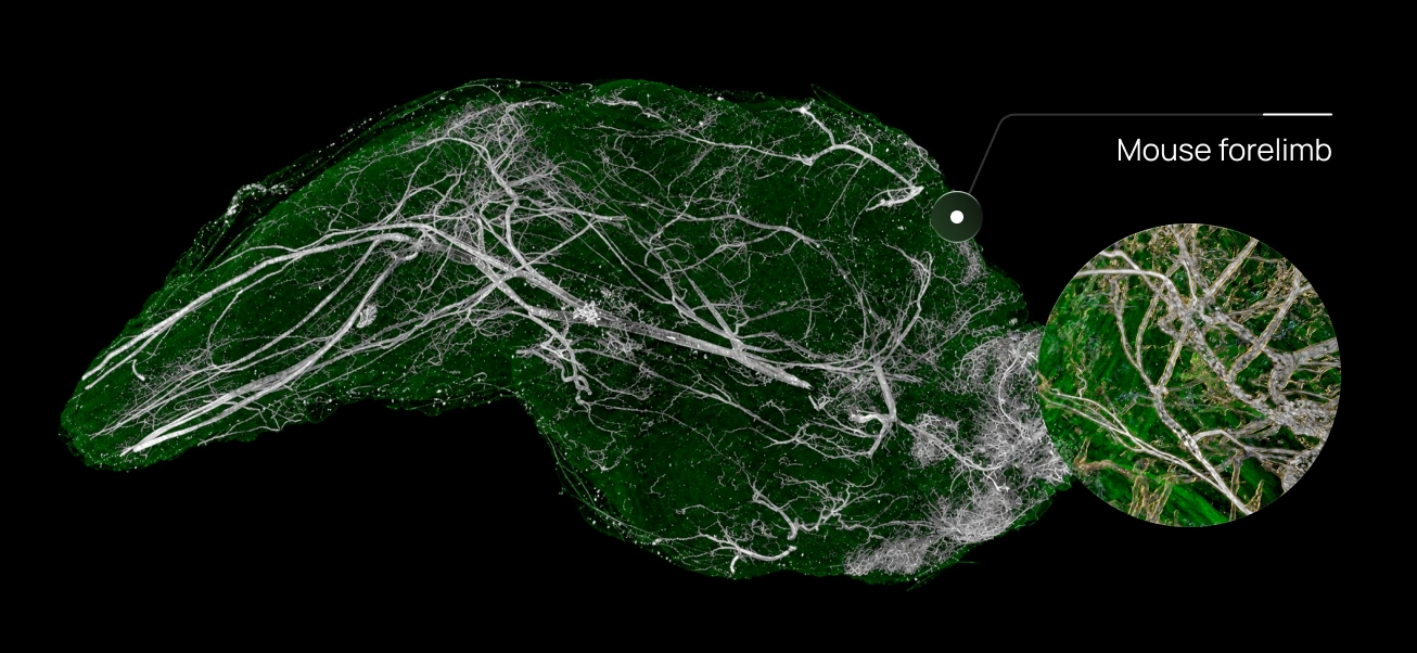
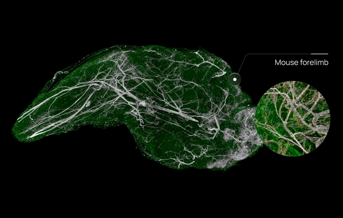
Cleared mouse forelimb imaged by the LiT XL2 light-sheet system

Cleared organs & tissues

Mouse forelimb vasculature with magnified detailMouse forelimb vasculature with magnified detail

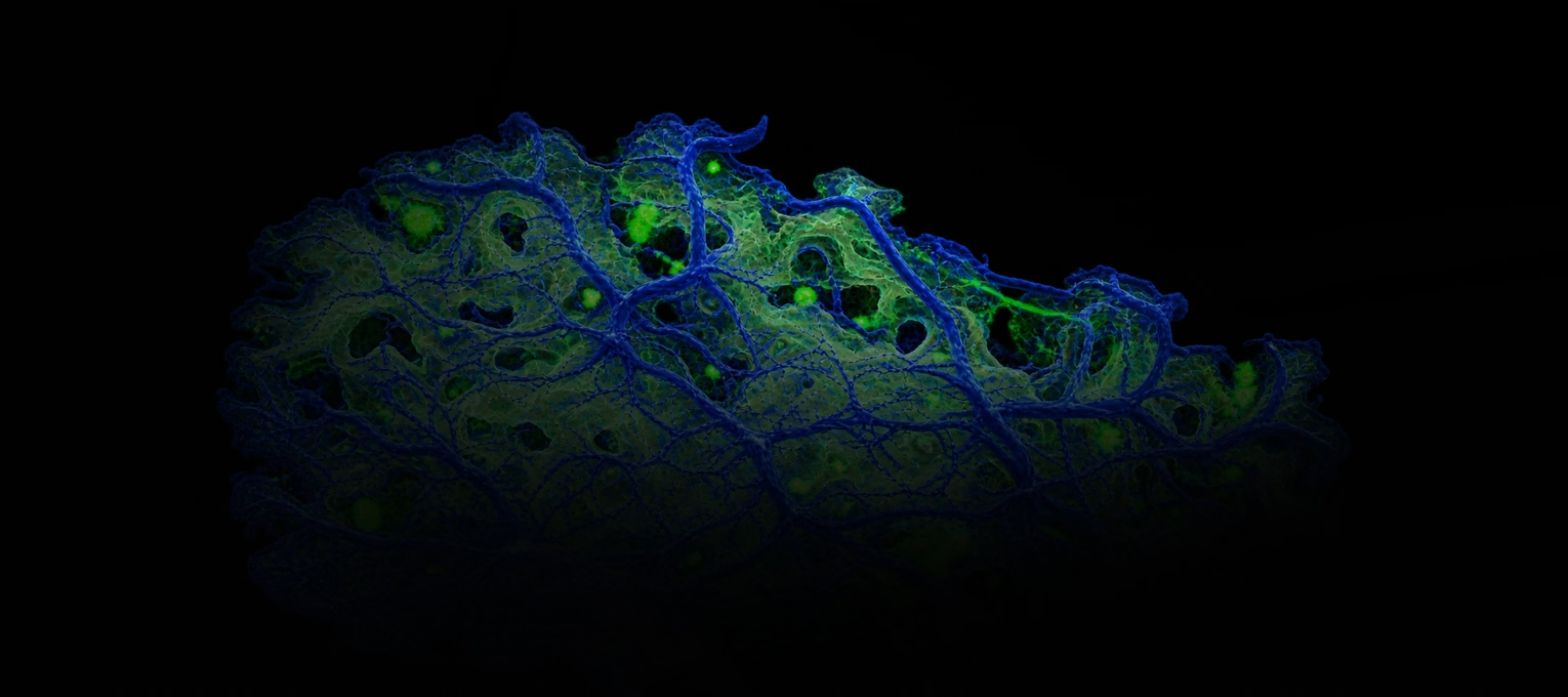
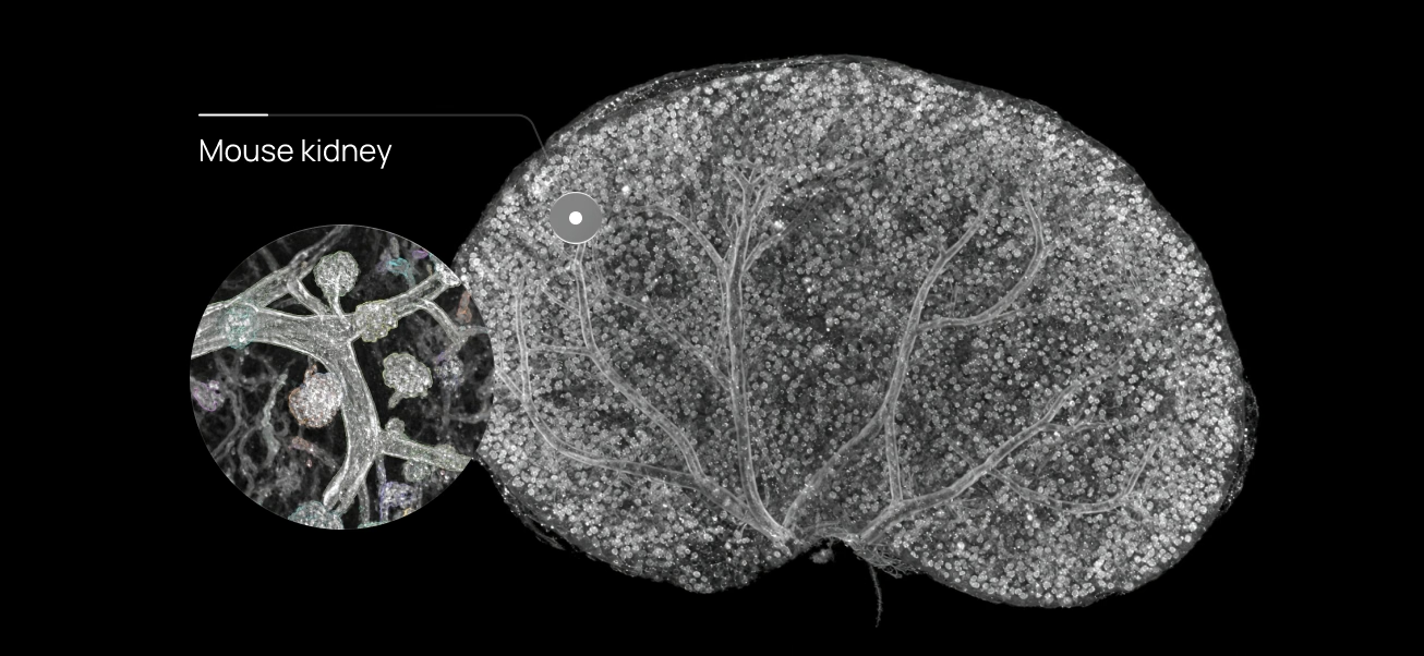
Whole mouse kidney imaging with glomeruli quantification

Cleared organs & tissues

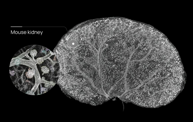
Mouse kidney rendering with magnified detailMouse kidney rendering with magnified detail

From acquisition to analysis-ready 3D data

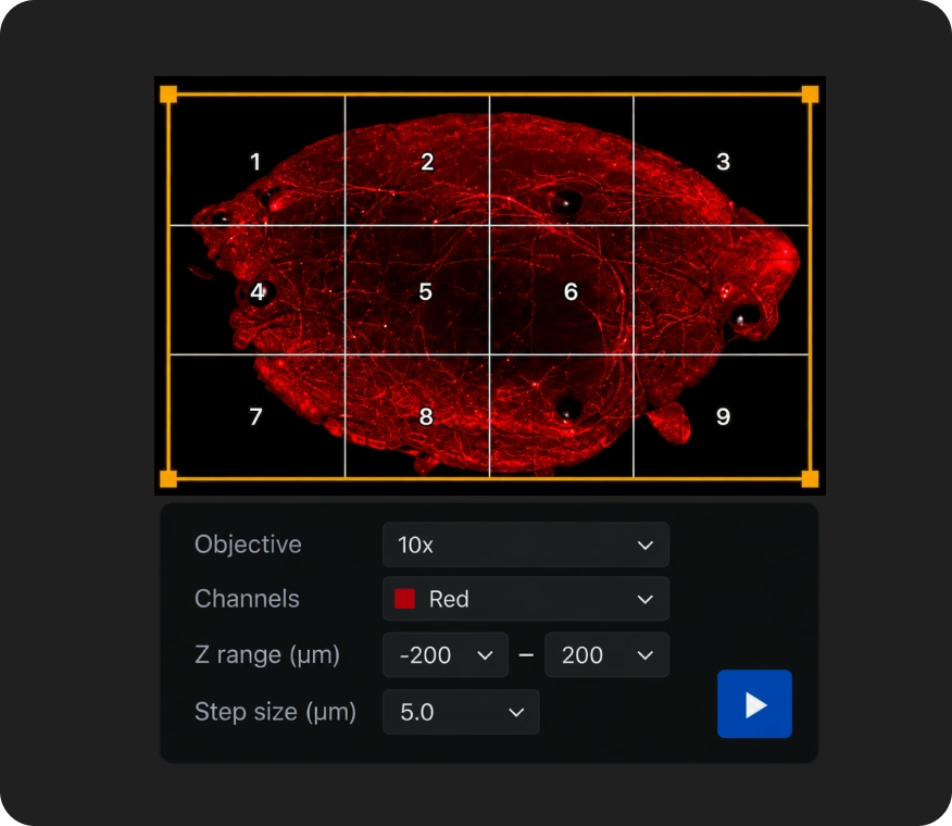
LiTScan acquisition controls over a red tissue scan
Acquire
Control acquisition and imaging.
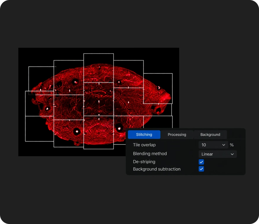
LiTScan processing controls over a red tissue scan
Process
Stitching, de-striping and background subtraction.
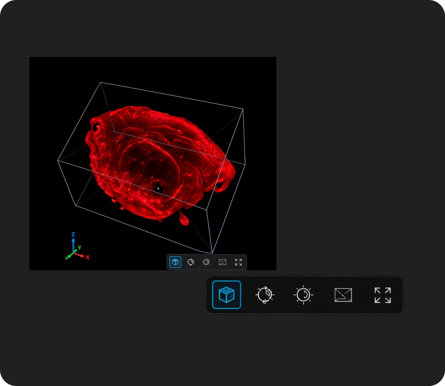
Three-dimensional rendering of a red tissue scan
Render
Build and visualize 3D volumes.
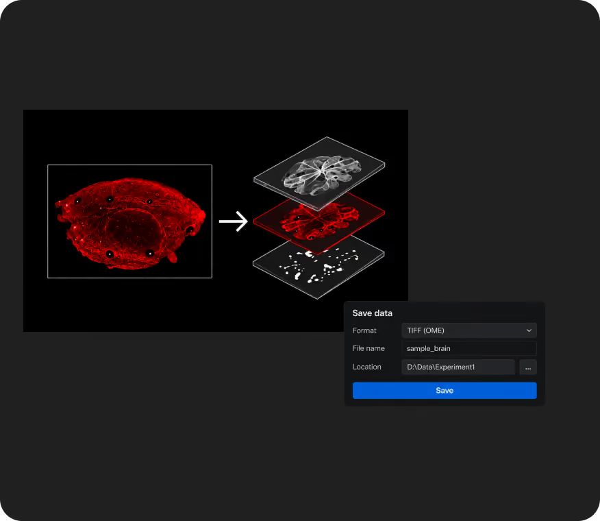
LiTScan export settings for a red tissue scan
Export
Prepare data for connected analysis tools.
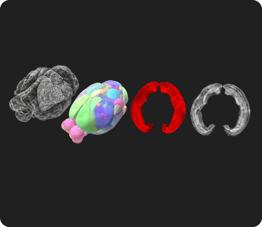
Four 3D tissue analysis visualizations
Analyze
Render in 3D or export to Imaris or Amira
Specifications

System specifications

Confirmed system parameters. Settings from individual published examples are not system specifications.

Objectives:4× / 5× / 10× / 16× / 25×
RI correction:Adjustable, 1.33–1.58
Illumination:Four-side Line Bessel Sheet illumination
Sample handling:Reusable, exchangeable sample bath;Holders for different sample sizes;Multiple mounting configurations
Sample size:2.5 cm × 2.5 cm × 2 cm, or 6.3 cm × 2.5 cm × 2 cmCustomization available for larger samples
Theoretical resolution:~310nm–887nm, wavelength dependent
System format:Compact benchtop design;No vibration table required under standard use;Vibration isolation may be useful at high magnification

How XL2 compares

Illumination approach

Dense-sample imaging

Refractive-index flexibility

Magnification range

Sample handling

System format

Processing workflow
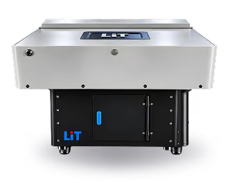
LiT XL2 microscope

XL2

Four-side Line Bessel Sheet illumination

Designed for more uniform illumination with reduced dark regions and striping

Adjustable RI correction from 1.33 to 1.58

Magnification range:4× / 5× / 10× / 16× / 25× objectives

Reusable sample baths and multiple holder configurations

Compact benchtop design

Acquisition, stitching, processing and 3D rendering supported through the LiT workflow
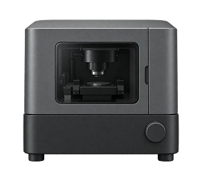
Typical alternative microscope

Typical alternatives

Commonly dual-side or multi-sheet illumination

Performance depends on illumination geometry and sample density

RI support varies by system

Objective options vary by platform

System-specific chambers and mounting workflows

Footprint and installation requirements vary

Often distributed across acquisition and downstream software tools

Support for complex imaging workflows

Setup & training

Installation, setup and system training.

Sample & imaging guidance

Clearing, mounting and acquisition support.

Data & analysis

Processing and analysis support.

We work alongside you at every stage, from setup to final results

Dr. Anna Keller
”
Research from the lab

Find the right system for your research