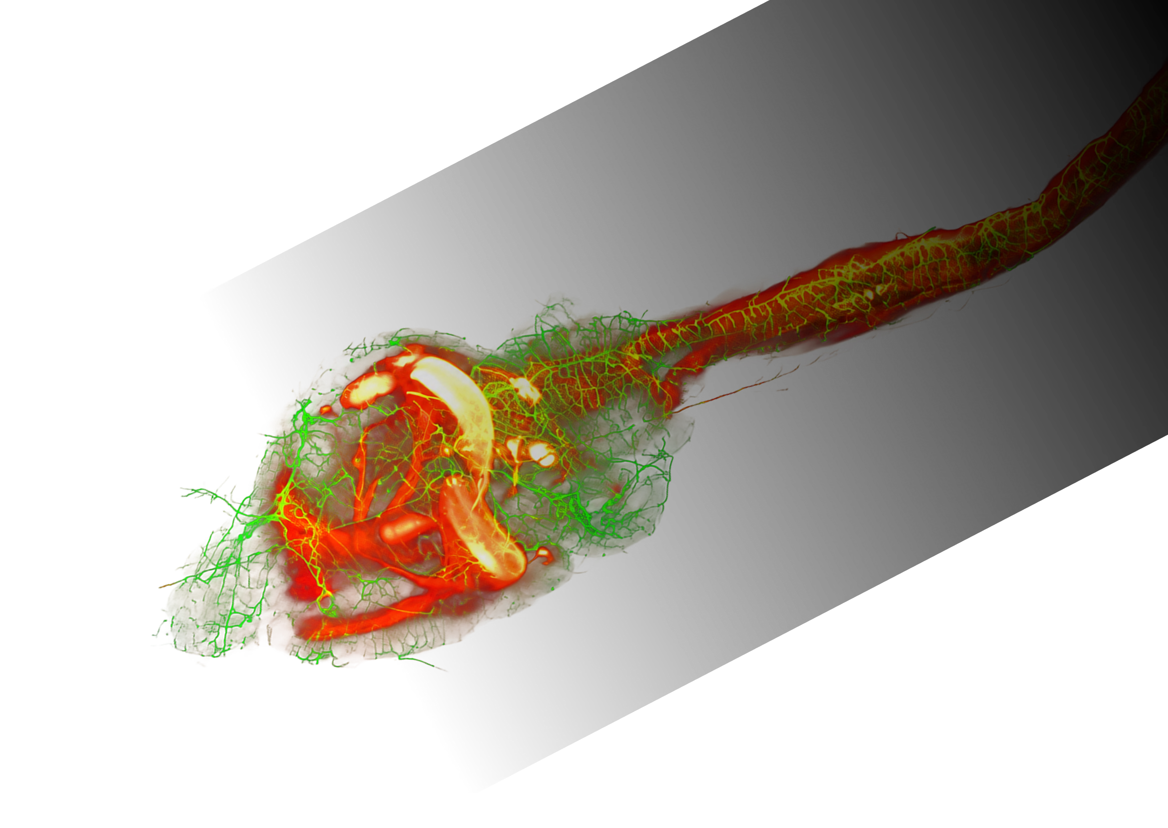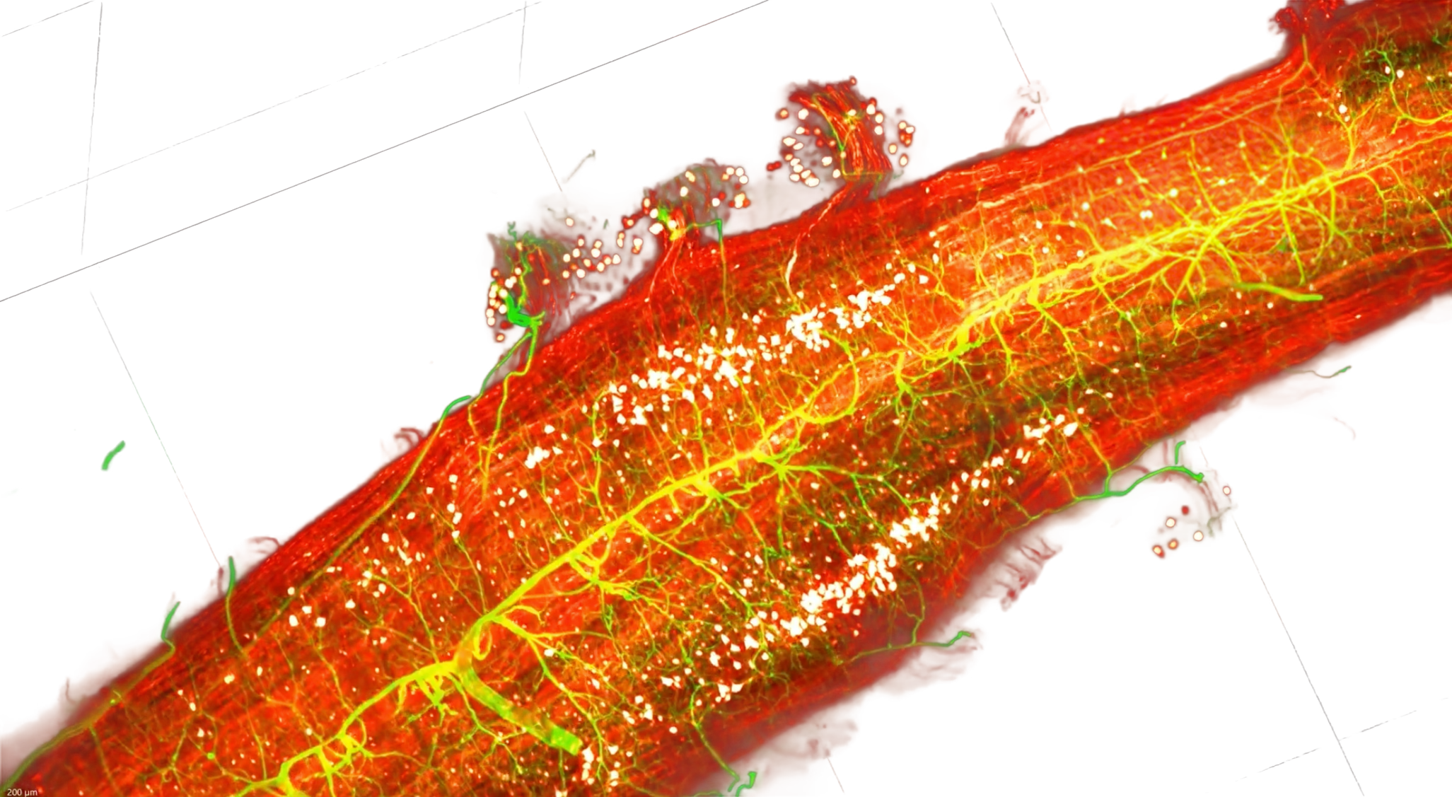

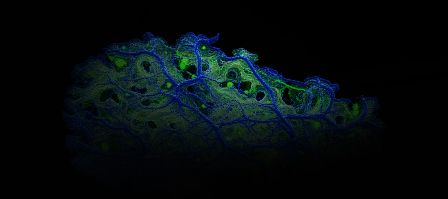
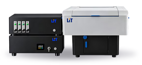
High-res imaging of intact cleared tissue

XL2 combines high-resolution volumetric imaging with flexible sample handling and an integrated acquisition-to-analysis workflow.

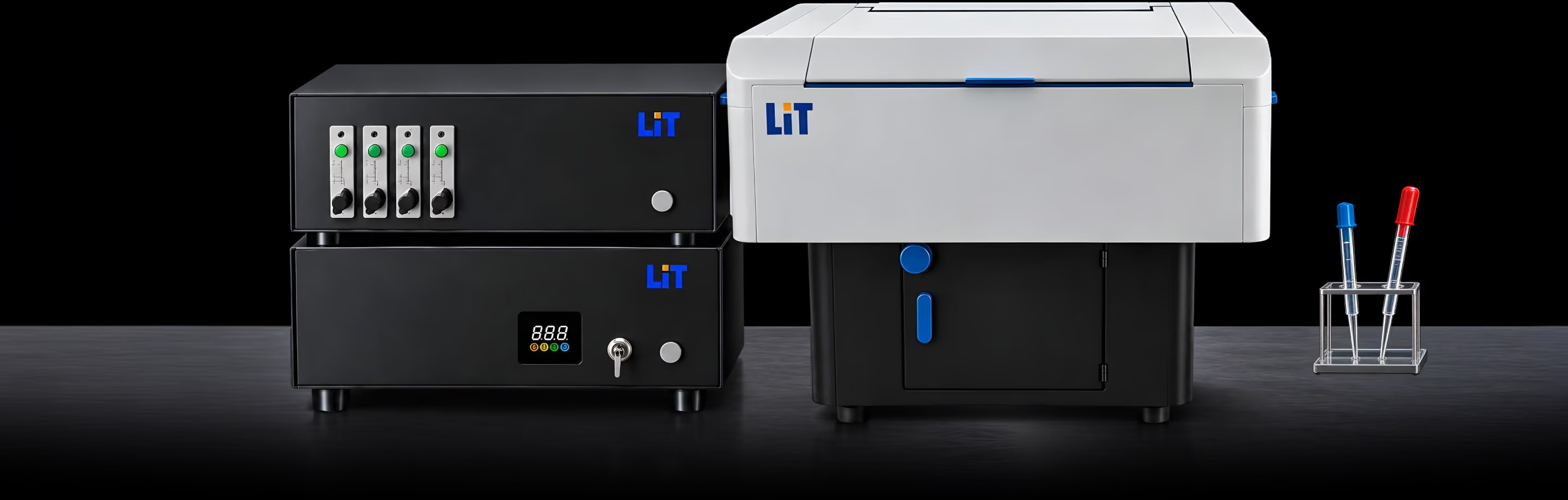
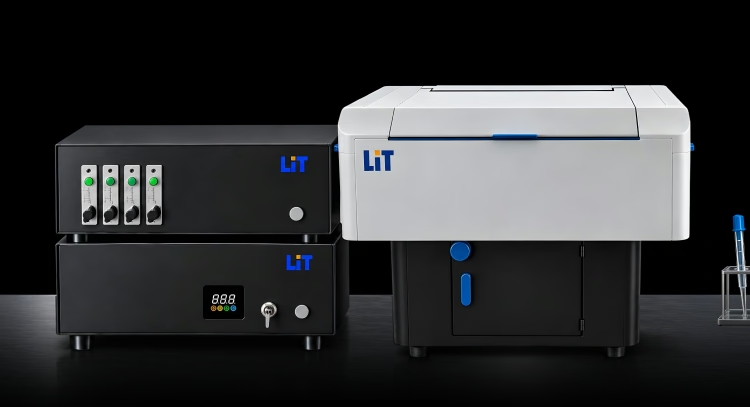
LiT XL2 microscope on a laboratory benchLiT XL2 microscope on a laboratory bench

Built for scale, automation and throughput

Large-volume imaging

Capture large biological volumes without losing the 3D context of the sample.

Built for automation

Reduce setup effort with automated calibration and chromatic correction.

Flexible across sample scales

Move from organoid arrays to whole-animal imaging within the same system.

Designed for throughput

Automated acquisition and processing help move more samples through the workflow.

Built for large-volume imaging

From organoid arrays to whole-animal specimens, UL combines scale, automation and flexible imaging modes in one system.

Large sample capacity
Up to 50 × 100 × 20 mm imaging volume
Flexible excitation
Single- or double-side illumination
Automatic calibration
Less manual setup between imaging sessions
Chromatic correction
Consistent multi-channel imaging across large volumes
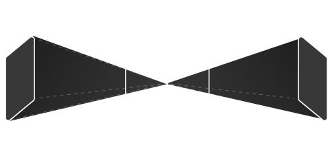
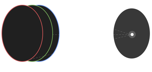
Flip imaging
Diagram of flip imaging for thick and complex specimens
Supports thick and complex specimens
All scales

Built to answer the questions your science needs to answer

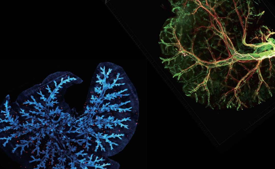
Microscopy visualization of cleared organs and tissues
Whole organs

Large-volume imaging of cleared organs and tissues for structural, cellular and spatial analysis

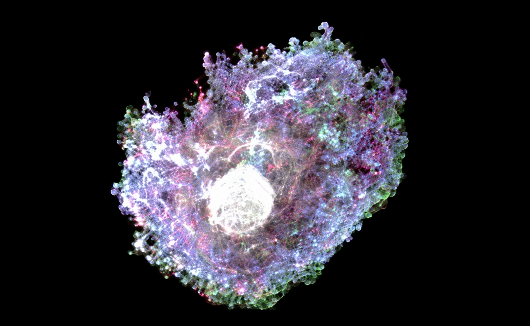
3D microscopy image of an organoid

Explore organoids in 3D, from overall structure to cellular detail.

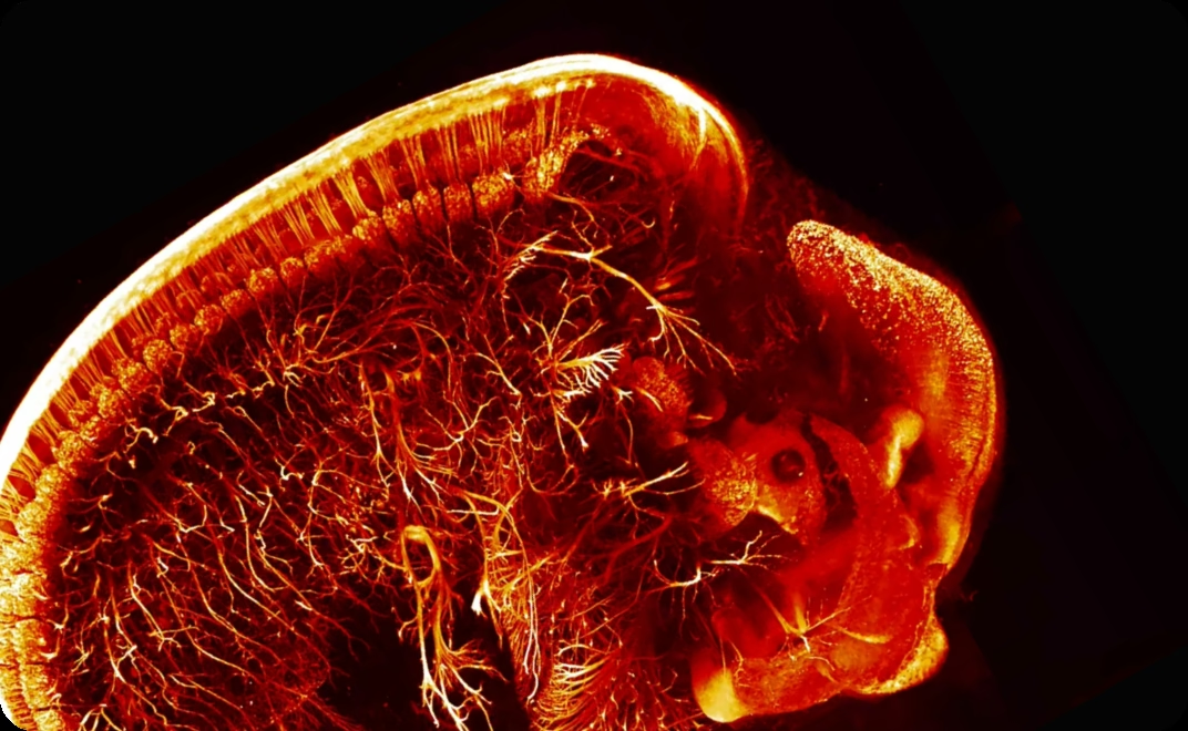
Microscopy image of a developing specimen

Visualize whole specimens across developmental stages to study morphology, growth and tissue organization

Microscopy visualization for cancer and biodistribution research

Visualize tumor burden and compound distribution across whole cleared specimens

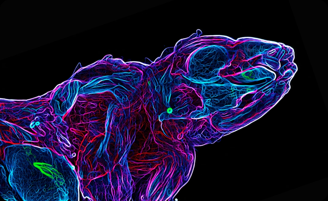
Whole-animal microscopy visualization

Image intact cleared animals to preserve spatial context across organs and systems

From acquisition to analysis-ready 3D data

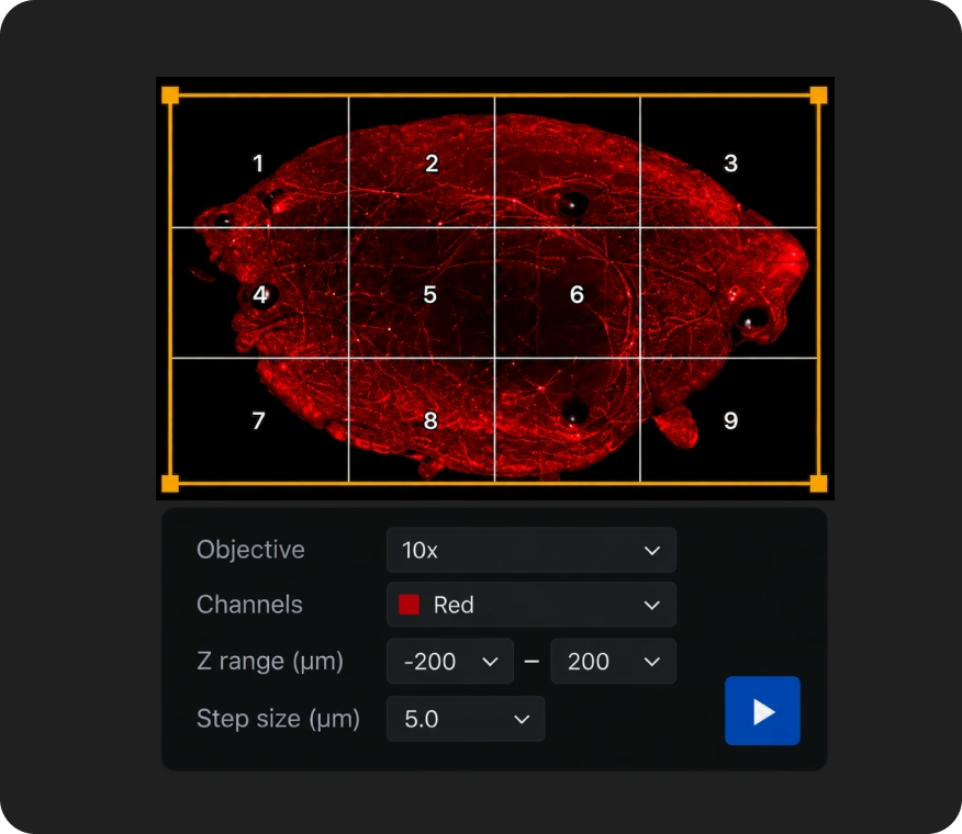
LiTScan acquisition controls over a red tissue scan
Acquire
Control acquisition and imaging.
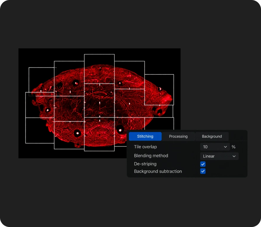
LiTScan processing controls over a red tissue scan
Process
Stitching, de-striping and background subtraction.
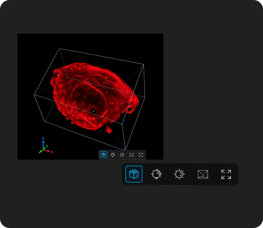
Three-dimensional rendering of a red tissue scan
Render
Build and visualize 3D volumes.
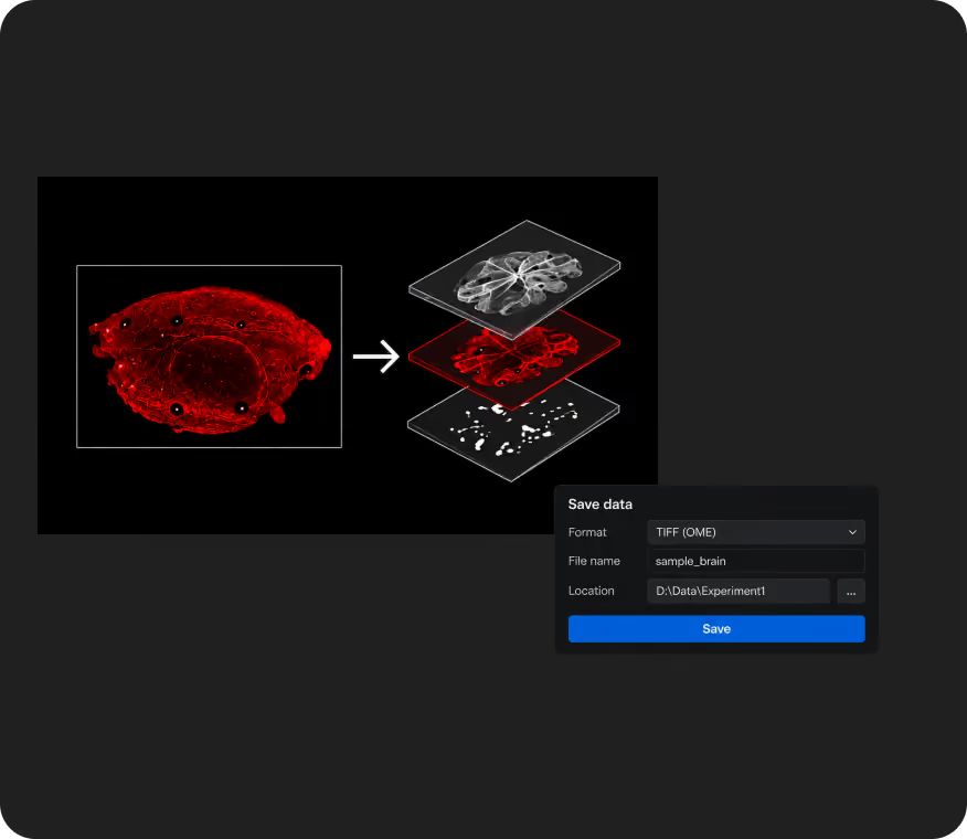
LiTScan export settings for a red tissue scan
Export
Prepare data for connected analysis tools.
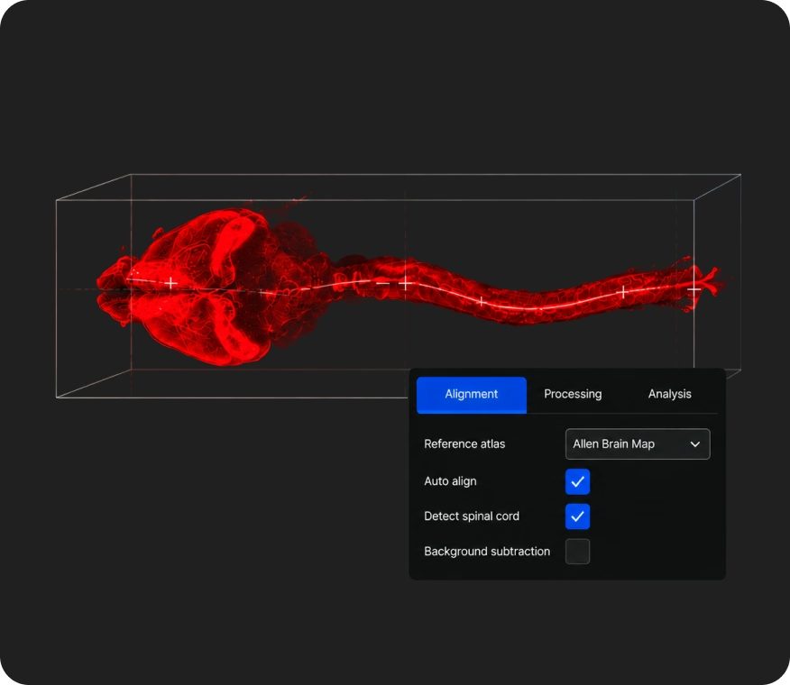
LITScan AiBot analysis visualization
LITScan AiBot
Automated brain and spinal cord alignment to Allen Brain Map. Automates processing and analysis workflows
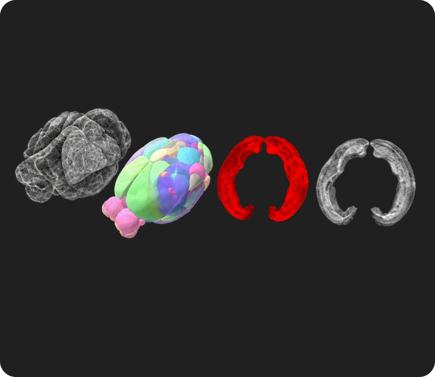
Four 3D tissue analysis visualizations
Analyze
Render in 3D or export to Imaris or Amira

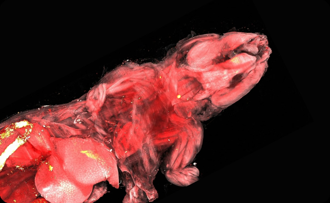
Whole-mouse imaging example

A large-volume workflow demonstrated on a 30 × 86 × 18 mm whole-mouse sample using a 5× objective

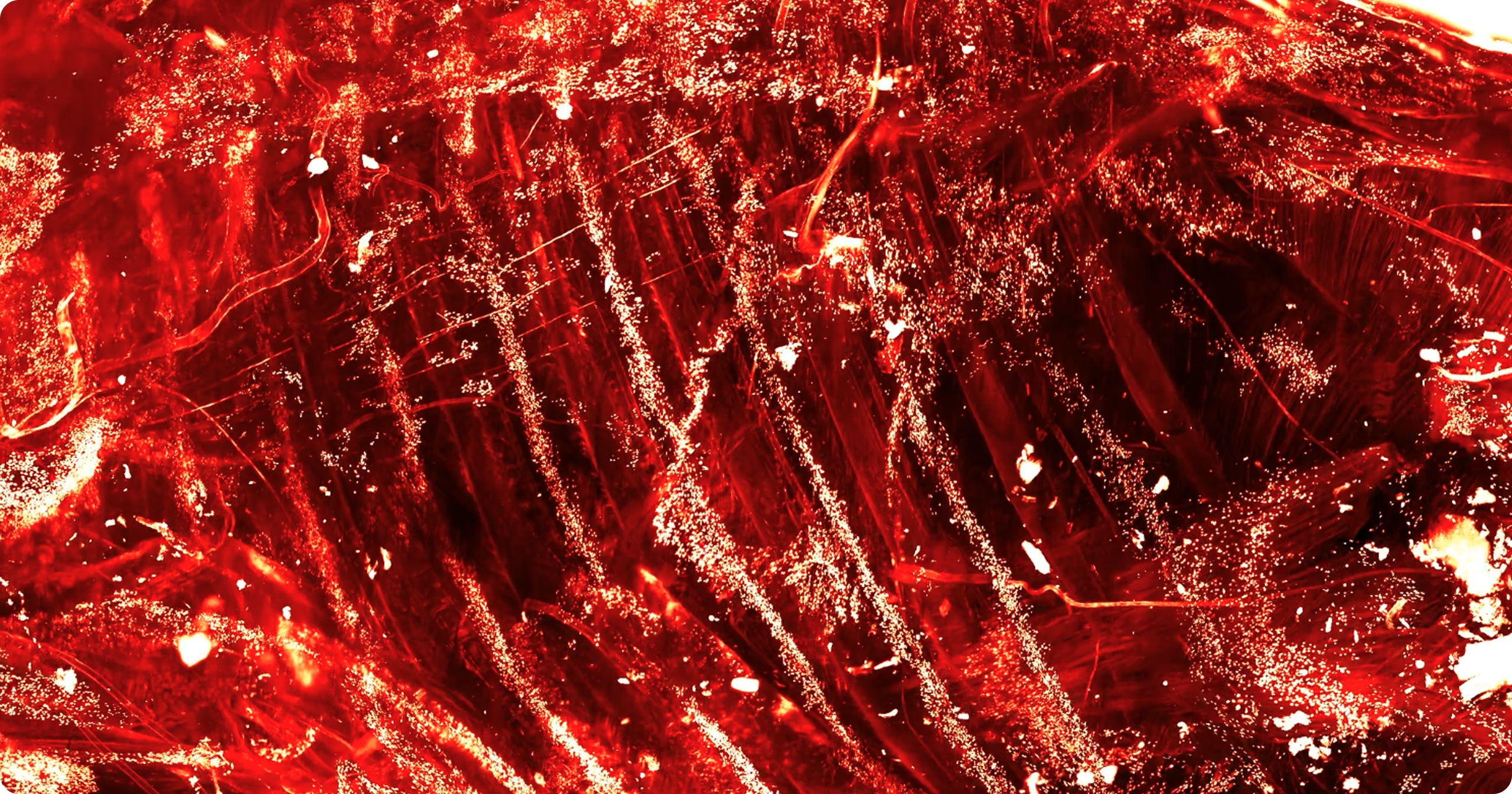
Red fluorescence microscopy image of a whole-mouse sample

49 x 90 x 40 mm

Maximum sample size

2.5x 2.5 x 2.5 mm

Single field of view

2.6 × 2.6 × 10 µm

Acquired voxel size

Specifications

System specifications

Confirmed UL parameters. Resolution is objective- and application-dependent.

Maximum imaging volume: 49 x 90 x 40mm (with flip z)
RI correction: Automatic
Chromatic correction: Automatic
Excitation: Single-side / double-side
Thick-sample imaging: Flip imaging
Sample finding: SmartScan
Software: LitScan
Standard objectives
2×NA 0.1Air
5×NA 0.35Air
10×NA 0.6Multi-immersion
25×NA 1.0Multi-immersion
Sample handling
Multiple holder designs
Enhanced corrosion resistance
Solid clamping
Easy flipping

How UL compares

Maximum sample volume

System alignment & calibration

Typical sample type

Illumination beams

Flip imaging for extra large samples

System objectives

Processing workflow

LiT UL microscope

UL

49x90x40mm (with flip z)

Automatic calibration and chromatic correction

Organoids to whole-animal imaging

Single- and double-side excitation

Flip imaging supported

2× / 5× / 10× / 25×

Acquisition, processing and large-volume data workflow through LiT through LiTScan software
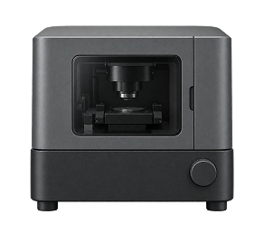
Typical alternative microscope

Typical alternatives

Maximum volume varies by platform

Level of automation varies by system

Often optimized for narrower sample ranges

Configuration varies by platform

Multi-orientation workflows vary

Objective range varies by platform

Often distributed across separate tools

Support for complex imaging workflows

Setup & training

Installation, setup and system training.

Sample & imaging guidance

Clearing, mounting and acquisition support.

Data & analysis

Processing and analysis support.

We work alongside you at every stage, from setup to final results

Dr. Anna Keller
”
Research from the lab

Find the right system for your research